|
RESEARCH DEVELOPMENT Kurt Grey Physiotherapist |
Seven Methods for Measuring the Treated Area |
|
Main Menu:
ULTRASOUND Research and Development:
Clinical guidance Tools and Tutorials:
MISCELLANEOUS:
LINKS Contact:
Downloads:
Latest update : 30-11-2011
|
Grey, K: ”Surface Areas in Ultrasound Therapy. Evaluation of Seven Measurements for Measuring the Treated Skin Area in Physiotherapeutic Application of Ultrasound.” Scand J Rehab Med, 25: 11-15, 1993. (Original paper, peer-reviewed).
ABSTRACT. Recent articles on ultrasound (US) do not contain information about precise dosage, thus risking a nonvalid conclusion of possible effects. To ensure safer standards in treatment time, the present study compared 7 methods for the calculation of areas. Measurement by eye had a median relative difference (MRD) from the true reading of 22% (lower quartile(l.q.)=-14%, upper quartile(u.q)=+46%), with large intraindividual variation, the span of MRD being ‑46% to +53%. Measuring with the transducer showed a systematic error: MRD was ‑48% (l.q.=-55%, u.q.=-35%), which is unacceptable. Areas were measured with satisfactory accuracy with 5 methods that require little training and use accessories, some of which are inexpensive. Plates with ellipses of known size had a MRD of 0.1%, (l.q.=-3%, u.q.=+2%), while rulers showed MRD of ‑2%, (l.q.=‑0.6%, u.q.=+5%) both on selected material. The most accurate and versatile instruments were a planimeter (MRD=-0.1%; l.q.=‑0.6%; u.q.=+0.5%), a Mettler weighing apparatus (MRD=3%; l.q.=2%; u.q.=6%), and a digitizer (MRD=-0.1%; l.q=‑0.4%; u.q=+0.3%).
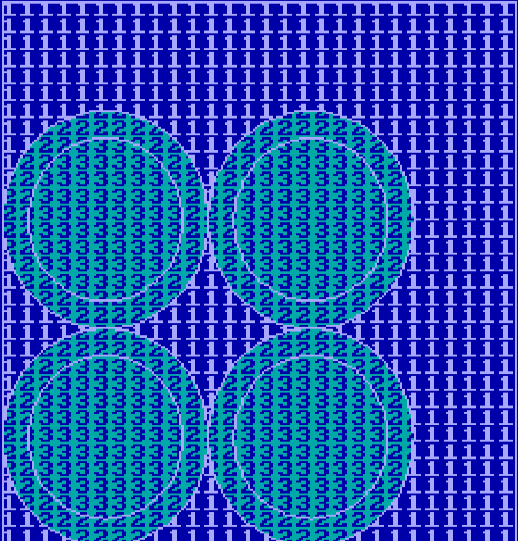
Fig 1. Measuring a rectangular lesion by the ultrasound transducer head (turquoise) tends to underestimate the area of the lesion. In the study above the underestimate was 48%. The transducer rarely fits the lesion exactly. The exceeding area is shown by dark blue covered with the numbers "1". The Beam area (ERA) (covered by the numbers "3") is usually smaller than the transducer housing ("2" and "3").
- 0 - |
